Technology
Given two images, that may or may not overlap, i2Align™
has two
inter-related jobs: to decide if the images do indeed overlap and, if
so, to compute an accurate mapping between the two
images. i2Align™ achieves this using a three-step
process: (1)
generating initial estimates of the inter-image transformation, (2)
refining the most promising initial estimates, and (3) deciding if any
of the resulting estimates align the same structures from the two
images. The earlier Dual-Bootstrap algorithm (GDB-ICP), IEEE
Trans. on Pattern Analysis and Machine Intelligence Nov 2007,
which we developed with colleagues and fellow students at Rensselaer
Polytechnic Institute, used matching of SIFT features to generate
initial estimates, a region growth and model selection algorithm to
refine the estimates, and a combination of stability, accuracy and
orientation-consistency measures to make the final decision. Each of
these components has been replaced with a proprietary new
technique in i2Align™, which is now more intelligent
and faster
than the Dual-Bootstrap algorithm.
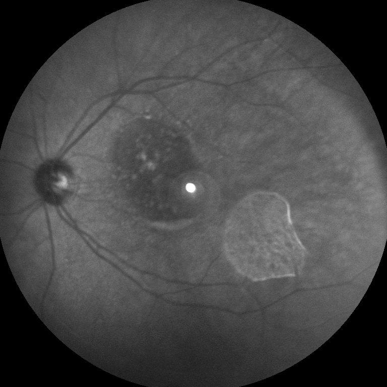
One example of the power of i2Align™ is the following gif animation of the alignment between a red-free fundus image and an ICG image.
Transformation Models
For fundus images, i2k Retina™ uses a quadratic transformation model which accurately accounts for the curvature of the retina when aligning color fundus images and fluoresceins. For working with non-fundus images, i2k Retina™ also offers similarity, affine, homography, planar (homography plus radial-lens distortion), and cylindrical (also with radial-lens distortion). See . These are accurate enough to produce well-aligned images in a wide variety of situations. Users who would like to experiment on their own have a choice of many other transformation models in addition to the quadratic model.
Automatic Masking
Due to the physical set-up of the image acquisition process, some non-photographic and most retinal fundus images contain an outer region that is outside the true image content. Automatically cropping this area is needed for clean montages, and it aids in the registration process, especially for low overlap images. When this exterior region is all black or all white, the masking process is trivial, but it can become considerably harder for poor quality images (e.g. images scanned from slides). By default, i2k Retina™ computes a mask for retinal fundus images, and includes an option to do this for non-fundus images.
Uneven Illumination and Vignetting Correction
Some photographic lenses and and most retinal fundus cameras produce
images that tend to be darker on the image periphery than near the
image center. For photographic lenses this is due to "vignetting",
while for fundus images it is caused by uneven illumination. In
either case, good quality montages and better overall appearance may
be obtained if this can be automatically corrected.
DualAlign™ has developed new algorithms for automatically correcting
vignetting and uneven illumination both in a single image and across
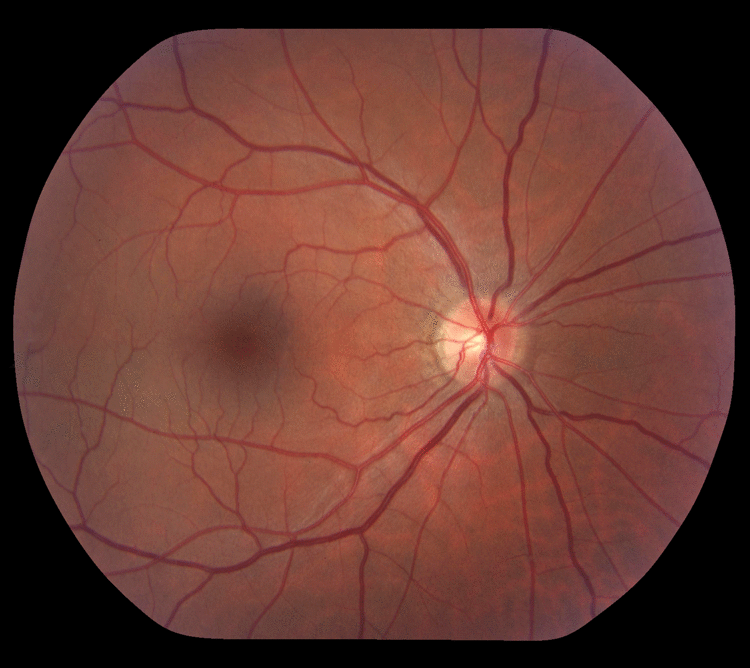
multiple images. These are applied, if desired by the user, in the retinal image montaging process of i2k Retina™. Below is an example of the effect of this correction process on a single image. Notice the much brighter image periperhy in the corrected image.
Seam Selection
i2k Retina™ include algorithms that select seams and blending regions between images based on a detailed model of the image misalignments and color differences that remain after illumination correction.
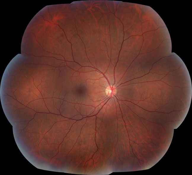
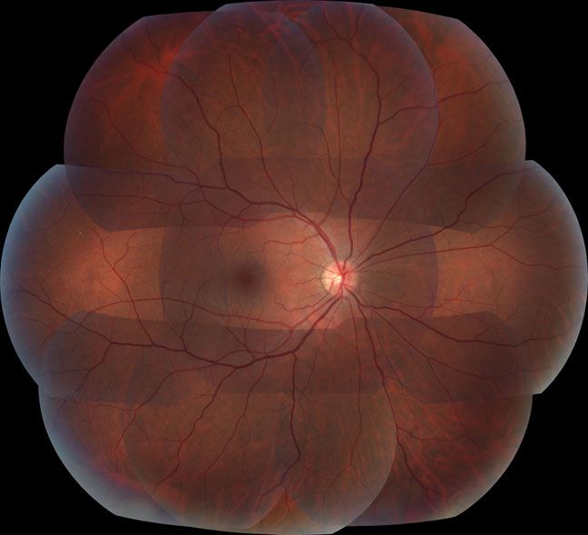
The following two montages shows the effects of using i2k Retina's tools for illumination correction and seam selection (top) and not using them (bottom). Users of i2k Retina™ can create either montage for themselves after loading the images with just a few mouse clicks.
|